The contest honors the best small-scale scientific photography of the year.
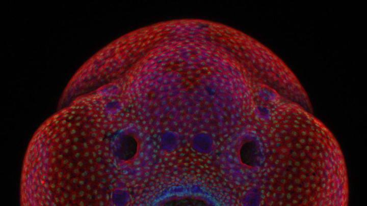
Each year, Nikon honors the best scientific photography on a micro scale with theNikon Small World contest, awarding the most impressive images taken through a microscope with up to $3000 towards new camera equipment. The top images will also be part of a national museum tour. This year’s winning image—taken by Oscar Ruiz of the University of Texas MD Anderson Cancer Center—is of a 4-day-old zebrafish embryo. Ruiz is using timelapse photography to study the development of the zebrafish face, hoping to gain insight into the cellular changes involved in facial deformities. The top 11 winning images can be seen below, but check out more winning images on Nikon’s Instagram, and stay tuned for the announcement of this year’s video winners on December 7.

1. A 4-day-old zebrafish embryo photographed by Dr. Oscar Ruiz, The University of Texas MD Anderson Cancer Center, Houston, Texas, USA. Ruiz explains how timelapse photography can illuminate the science of facial deformities:

2. A polished slab of Teepee Canyon agate photographed by Douglas L. Moore, University of Wisconsin - Stevens Point, Museum of Natural History, Stevens Point, Wisconsin, USA

3. A culture of neurons (stained green) derived from human skin cells, and Schwann cells, a second type of brain cell (stained red) photographed by Rebecca Nutbrown, University of Oxford, Nuffield Department of Clinical Neurosciences, Oxford, United Kingdom

4. Butterfly proboscis photographed by Jochen Schroeder, Chiang Mai, Thailand

5. An image of the front foot (tarsus) of a male diving beetle by Dr. Igor Siwanowicz, Howard Hughes Medical Institute (HHMI), Janelia Research Campus, Ashburn, Virginia, USA

6. An image of air bubbles formed from melted ascorbic acid crystals by Marek Mis, Marek Mis Photography, Suwalki, Podlaskie, Poland

7. Leaves of Selaginella (lesser club moss) photographed by Dr. David Maitland, Feltwell, United Kingdom

8. Wildflower stamens photographed by Samuel Silberman, Monoson Yahud, Israel

9. Espresso coffee crystals photographed by Vin Kitayama and Sanae Kitayama, Vinsanchi Art Museum Azumino, Azumino, Nagano, Japan

10. Frontonia (showing ingested food, cilia, mouth and trichocysts) photographed by Rogelio Moreno Gill, Panama, Panama

11. Scales of a butterfly wing underside (Vanessa atalanta) photographed by Francis Sneyers, Brecht, Belgium
