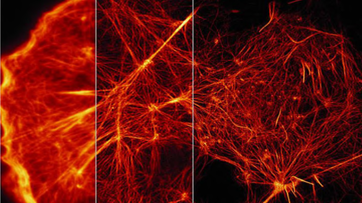
Since Robert Hooke first discovered the cell in 1665, scientists have been peering through microscopes in an effort to learn more about these basic units of life. In the 350 years since, technological advances have allowed us to look closer at how cells function, but not everything we know comes from first-hand observations. Some cell activity—like the hundreds of tiny bubbles surfacing on a cell at any given time—moves too fast for the human eye to register, even when looking through the strongest microscope. The details of this molecular-level action have been inferred.
But now scientists have found a new way to capture cell life in unprecedented detail, as they demonstrated last week in a series of photographs published in the journal Science that reveal the inner workings of cells never before seen by the human eye. The breakthrough was made possible by a technique called structured illumination microscopy, or SIM, which is used in filmmaking.
Two years ago, Harvard cell biologist Thomas Kirchhausen attended a lecture by Eric Betzig of the Howard Hughes Medical Institute’s Janelia Research Campus on the use of SIM to study cells. Betzig's previous work included developing a technique of high-resolution microscopy that uses fluorescent molecules to highlight parts of the cell. (He shared the 2014 Nobel Prize in chemistry for this work.)
The problem with this method is that it exposes the cells to light that’s more intense than what they’re equipped to handle, which ends up harming and sometimes even vaporizing them. But SIM is gentler, capturing images of live cells much faster while using less light.
Kirchhausen thought it might be possible to use SIM at the molecular level to capture cell activity. He and Betzig subsequently collaborated with researchers in China and the U.S., and the result was this set of groundbreaking images. Check out an example in the video below, which uses magenta and green flourescent molecules to highlight the proteins actin (magenta) and myosin (green) working together to form the network of filaments necessary to cell movement.
[h/t: MIT Technology Review]
