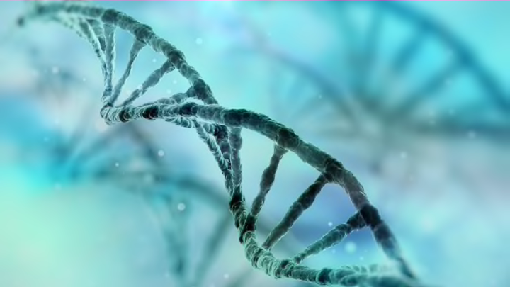
Picture a strand of DNA and the image you see will likely be similar to the artist’s rendering above. The iconic twisted ladder, or double-helix structure, was first revealed in a photo captured by Rosalind Franklin in the 1950s, but this popular visualization only tells part of the story of DNA. In the video below, It’s Okay to Be Smart explains a more accurate way to imagine the blueprints of life.
Even with sophisticated lab equipment, DNA isn’t easy to study. That’s because a strand of the stuff is just 2 nanometers wide, which is smaller than a wavelength of light. Researchers can use electron microscopes to observe the genetic material or x-rays like Rosalind Franklin did, but even these tools paint a flawed picture. The best method scientists have come up with to visualize DNA as it exists inside our cells is computer modeling.
By rendering a 3D image of a genome on a computer, we can see that DNA isn’t just a bunch of free-floating squiggles. Most of the time the strands sit tightly wound in a well-organized web inside the nucleus. These balls of genes are efficient, packing 2 meters of DNA into a space just 10 millionths of a meter across. So if you ever see a giant sculpture inspired by an elegant double-helix structure, imagine it folded into a space smaller than a shoe box to get closer to the truth.
[h/t It’s Okay to Be Smart]
