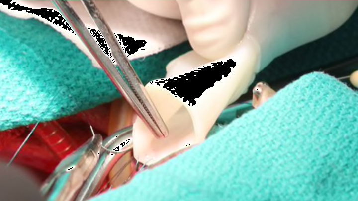
Welcome to the future. Scientists have created arteries that can be safely implanted and continue growing in their hosts. They published a report of their progress today, September 28, in the journal Nature Communications.
Transplanted organs and tissue face several major obstacles to success. First, there’s ensuring the transplant is right and safe for the recipient. Then there’s the possibility that the recipient’s body will reject the new part. Finally, there’s the need for the implanted materials to cooperate with the cells around them, to grow and work together. Scientists have made major headway on the first two issues over the last few decades. But when it comes to coaxing transplanted parts to grow, we’re really just getting started.
Growth is especially important—and hard to produce—in blood vessel transplants. Scientists have found ways to make it happen, but they involve growing new vessels in the lab from scratch, using each patient’s own cells. The customization process is expensive and time-intensive, which seriously limits its use.
So a team of researchers at the University of Minnesota set out to find another way. They essentially wanted to build a generic or base model of the pulmonary artery—one that could be kept on hand in a hospital and used as needed.
They started with sheep. The team took samples of sheep skin cells and mixed them with a clotting agent and calcium chloride to give them rigidity, then pumped them into a tube-shaped glass mold. As the cells took shape in the tubes, the researchers infused them with nutrient fluids to give them the shape and flexibility they would need. They then transferred the cells to a bioreactor for another five weeks of maturation and stretching.
Once the arteries had grown and stretched to the right size, the team rinsed them in chemicals that stripped out all the original skin cells, a process known as decellularization. All that remained were the newly grown structures themselves; the shapes of blood vessels, with none of the immune system–triggering cells.
The new arteries were then implanted in three 8-week-old lambs. The lambs were patched up, then monitored with regular ultrasound scans 8 weeks, 30 weeks, and 50 weeks after their surgery. After the last scan, the lambs were euthanized and their arteries removed and dissected.
The artificial blood vessels had fared incredibly well. Not only did the lambs’ bodies not reject the grafts, but they seemed to embrace them. The transplanted arteries entered the lambs’ bodies as scaffolds, essentially, yet by the time the animals reached young adulthood the scaffolds were filled and composed of their own cells. The blood vessels grew with their owners, serving them well.
Jeffrey Harold Lawson is a professor of vascular surgery at Duke University. "This appears to be very exciting work and continues to support the emerging field of vascular tissue engineering," Lawson, who was unaffiliated with the study, told mental_floss. "It is very exciting to see the vessels grow over time with the sheep and repopulate with the hosts' own cells. If work like this continues to make both preclinical and clinical progress, it could revolutionize the field of pediatric cardiac surgery and potentially avoid reoperative procedures for thousands of young children."
Know of something you think we should cover? Email us at tips@mentalfloss.com.
