10 Award-Winning Microscope Images That Blur Science and Art
By Abbey Stone

Some of the most vibrant, beautiful photographs taken this year weren’t captured with a camera, but with a microscope.
The Olympus BioScapes Digital Imaging Competition honors outstanding images and videos of life science subjects “shot” with a light microscope. Each year, nearly 2,500 still images and movies are submitted by scientists from over 70 countries. From those, 10 are chosen as winners.
Of this year’s crop of winners, the BioScapes website says, “The beauty, power and importance of science as portrayed by these incredible images and movies captivated this year’s panel of judges and is delighting viewers worldwide.”
Check out 2014’s 10 winners (and prepare for your world to be rocked):
1. Dr. William Lemon, Dr. Philipp Keller, Fernando Amat // HHMI Janelia Research Campus; Ashburn, Virginia
In their winning video (which can be seen at OlympusBioScapes.com), Lemon, Keller, and Amat show multiple views of Drosophila (a genus of small flies) embryonic development. The scientists recorded the embryo in 30-second intervals over a 24-hour period, beginning three hours after egg laying. At the end of the video, you can see the newly-hatched larva begging to crawl out of the frame.
2. Thomas Deerinck // National Center for Microscopy and Imaging Research, University of California; San Diego, California
At first glance, Deerinck’s image looks like a flower bed. In reality, it’s a rat brain cerebellum.
3. Dr. Igor Siwanowicz // HHMI Janelia Research Campus; Ashburn, Virginia
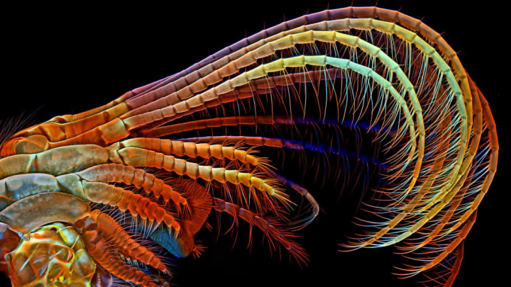
Dr. Siwanowicz’s third prize-winning image shows Crayola-hued barnacle appendages that sweep plankton and other food into the barnacle’s shell.
4. Dr. Csaba Pintér // Keszthely, Hungary
Phyllobius roboretanus weevils continue the circle of life.
5. Madelyn May // Hano, New Hampshire
More rat brains! Madelyn May scored fifth place with her confocal microscopy image of a rat brain cerebral cortex. The cell nuclei are cyan (greenish-blueish), the astrocytes are yellow, and the blood vessels are red.
6. Dr. David Johnston // Southampton General Hospital Biomedical Imaging Unit; Southampton, United Kingdom
Dr. Johnson’s image shows a magelonid polychaete worm larva found in a plankton sample collected in Southampton Water off the south coast of the United Kingdom. The actual specimen was approximately 2mm long.
7. Oleksandr Holovachov// Ekuddsvagen, Sweden
Holovachov brilliantly captures a butter daisy (Melampodium divaricatum) flower at 2x magnification using fluorescence.
8. Dr. Matthew S. Lehnert, Ashley L. Lash // Kent State University at Stark; North Canton, Ohio
This spiky proboscis (mouthparts) belongs to a vampire moth (Calyptra thalictri) captured by Jennifer Zaspel in Russia. The image shows the dorsal legulae, tearing hooks, and erectile barbs that helps the moth take in fruit juices and mammal blood while eating.
9. Dr. Igor Siwanowicz // HHMI Janelia Research Campus; Ashburn, Virginia
The gear-like trochanters (the part of the femur that connects to the hip bone) shown in Dr. Siwanowicz’s image help propel the green boneheaded planthopper (Acanalonia conica) at a staggering acceleration of 500 times the force of gravity. This image also reveals that gears, until recently thought one a human invention, in fact exist in nature.
10. Dr. Philipp Keller, Fernando Amat, and Misha Ahrens // HHMI Janelia Research Campus; Ashburn, Virginia
First-prize winner Dr. Keller also took home tenth place for his video of neural activity in an entire zebrafish brain in vivo. The 33-second clip marks the first time single-neuron activity in the brain of a living vertebrate has been captured in such exhaustive detail.
All images courtesy 2014 Olympus BioScapes Digital Imaging Competition®; www.OlympusBioScapes.com