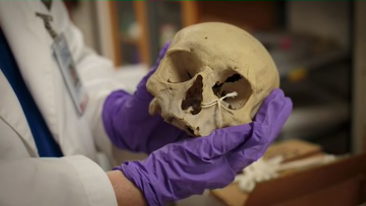
On a cart in Anna Dhody’s office sits a small, innocuous box marked “caramel Danish rolls.” Open it up, though, and you won’t find a pastry; instead, there’s a human skull nestled inside. Nearby, there’s another cardboard box—this one labeled “brain slices”—and on the bookshelf sits a jar of dried human skin.
The presence of these items might seem pretty weird—if not alarming—under typical circumstances, but this is not a typical office. Dhody is a forensic anthropologist and the curator of Philadelphia’s Mütter Museum, which houses anatomical specimens, models, and instruments from medical history. Visitors to the museum—which was founded by the College of Physicians of Philadelphia in 1858 from the collection of surgeon Thomas Mütter—can see the tumor removed from President Grover Cleveland’s jaw, slices of Einstein’s brain, a plaster cast of conjoined twins Chang and Eng, a dermoid cyst, and the tallest skeleton on display in North America.
But there’s much more that the museum doesn’t have on display. Dhody took us on a tour to give us a peek at what the public doesn’t get to see. (For more on the life of Thomas Mütter and the museum’s history, pick up Cristin O'Keefe Aptowicz’s excellent book, Dr. Mütter’s Marvels: A True Tale of Intrigue and Innovation at the Dawn of Modern Medicine.)
1. An Iron Lung
Though we now associate the iron lung with polio, the device was originally invented for coal miners who had inhaled toxic gasses, according to Dhody. This particular iron lung, used in the 1950s, was an Emerson Negative Pressure Ventilator. “I love this piece—it’s amazing—but it’s so big,” Dhody says; she estimates that it weighs more than 800 pounds. The machine would fit someone over six feet tall; his head would stick out, while his body was inside the chamber. “Your whole respiratory system is under a little bit of negative pressure—that’s what fills [the lungs] up with air and that’s what you need to breathe,” Dhody says. “So the engine would generate artificial negative pressure inside the chamber and basically force your ribcage to move up and down and allow you to breathe.” If the power went out, nurses would manually operate a bellows at the end of the iron lung to keep the negative pressure going. Though the machines aren’t used much anymore, “as of 2008, there were 80 people in the world that still use iron lungs either full- or part-time,” Dhody says.
2. A Mystery Penis
Though the Mütter is known for its human remains, the museum also has a fair amount of animal remains, which are important for comparative anatomy purposes. Picking the favorite of that collection is easy. “I can tell you that it’s a penis," Dhody says. "What I can’t tell you for sure is what kind of animal it comes from.” Though the tag reads “horse,” Dhody’s friend—an equine facilitator who owns a horse farm and “knows her horse junk”—put that myth to rest.
The preserved member is huge—easily the length of a person's arm—and Dhody isn’t sure when it became part of the collection. “It has an F number, which means found number,” she says. “It was way before my time.” Based on x-rays of the baculum, or penis bone, taken by the Philadelphia Zoo, “we’re basically thinking that it’s from a larger sea mammal—a walrus, sea lion, or elephant seal,” Dhody says.
3. A Jar of Human Skin
In 2009, a young woman with dermatillomania—a mental disorder that creates a need to pick skin off the body—donated a jar of skin she had picked off of her feet to the museum. Dhody promptly put that jar on display. Fast forward to 2014: “She’s still picking, and I’m still taking it,” Dhody says of the new jar of skin, donated earlier this year, that sits on her bookshelf. “What’s interesting is that this seems to churn people’s stomachs more than the severed limbs and heads in jars. They see this and go ‘ahh!’ I don’t get it. It’s skin.” There was a good reason for taking the skin, which, by the way, smells like romano cheese. “It has huge educational impact,” Dhody says. “How many other ways do I have of showing a physical manifestation of a mental disorder?” (Thankfully, the young woman making these donations only picks from her feet; others with dermatillomania can disfigure themselves.) Dhody might eventually combine both donations into a single jar for display, but, she says, “I like having it in my office right now.”
4. A Replica of Chamberlen Forceps
In the 1600s, two Chamberlen family brothers, both named Peter, worked as surgeons and obstetricians and invented modern obstetrical forceps—a technology that the family kept secret for a century. “The whole concept of intellectual property and who owns rights—it’s not just computer stuff,” Dhody says. “For a hundred years, this family dominated the field of obstetrics in Europe. They could command up to $10,000 [in the money of that time] for one birth all because of these two pieces of metal.” The Mütter’s pair of Chamberlen forceps is a metal replica that was made a couple of hundred years ago.
The design of modern forceps hasn’t changed much since the Chamberlens. “The one thing that is different is that those blades can come apart so you can insert one at a time into the vaginal canal, whereas [with this tool], they’re together,” Dhody says. “It’s amazing that this technology still exists. Forceps are going out of favor, but they are still used—not as much in America, but other parts of the world. It’s good when a baby is just stuck in there. If you are a practitioner who is skilled in the use of forceps, it’s safe. You know, there are risks involved, but there are more risks if the baby is stuck.”
5. A Pessary
“If you think about the skeletal structure of the human body—especially the lower abdominal area—there is no skeletal support structure for certain internal organs, including the uterus,” Dhody says. “Often times as a woman ages, or when she’s had a lot of children, the muscles, ligaments, and tendons will weaken and the uterus falls out of position; it can go down and through the vagina.” These days, doctors would probably opt to do a hysterectomy, but before that option was available, women “would insert an object up the vagina to kind of wedge it and keep that uterus from falling out,” Dhody says. The devices were called pessaries, and the museum has hundreds of them, including this one from the 19th century, which the curator finds particularly creepy because of the springs. “You can take it out, clean it, put it back in,” Dhody says. “You can still see these today—it’s a medical tool. But now they’re made out of surgical grade plastic. If you’re living in an area where surgery isn’t an option or if you have certain religious objections to that, then a pessary is a good bet.”
6. A Blood Circulator
This device, from the early 20th century, claimed to do “for internal organs what exercise will do for the limbs” as well as alleviate cold symptoms. “It was a suction thing,” Dhody says. “You’d push it against the body and it vibrates.” The instruction manual is full of photos of a presumed doctor placing the circulator on various parts of a woman’s body and cranking the handle, which the manufacturer claimed would create suction and increase blood flow to a particular area.
7. A Skull with Artificial Cranial Deformation
Dhody rediscovered this skull in the museum’s mobile storage. “I came across these boxes that said ‘caramel Danish rolls,’” she says, “and I was like, ‘What are the chances that these actually have caramel Danishes in them? Very little.’" She adds jokingly, "that happens a lot—it’s never a Danish. Sometimes you have enough skulls, and you just want a Danish.”
The skull, which has been artificially deformed, comes from Peru; Dhody guesses it’s from around the 1800s. “People in Peru practiced artificial deformation for hundreds and hundreds of years,” she says. “Up until about the 20th century, [there were] remote parts that were practicing it.”
8. Dhody’s Husband’s Gallbladder
“We have a friends and family plan at the Mütter,” Dhody says. “It’s basically acknowledged that if you work here, or if you are associated with anyone who works here, and you lose any body part for any reason, we have dibs.” So when her husband had his gallbladder removed, Dhody jumped at the chance to both watch the operation and make it part of the collection. “Unfortunately, it looks perfectly healthy,” she says. “I guarantee you, it was not when it was removed." Gallstones, she says, block the bile duct and cause inflammation, pain, and vomiting until they’re removed. Patients will get big stones—which you can see in the photo below—or microstones, which look like sludge and more easily block the duct.
Dhody's husband had microstones, so his gallbladder had to come out. To remove the gallbladder, surgeons made five small incisions—including one through the belly button—and inserted laparoscopic tools. “Then, they seal the gallbladder in, like, a tiny little body bag and tug it out,” Dhody says. Now, her husband’s gallbladder sits preserved in alcohol in the Mütter’s wet room, where the museum’s on-site conservation also takes place.
9. Intestinal Specimen
The wet specimens are housed in a climate controlled room where the air is exchanged six to eight times every hour, with redundant units to ensure it’s always the proper temperature. The majority of the Mütter’s specimens are preserved in alcohol, which doesn’t destroy DNA. “This doesn’t look too interesting because it’s just a intestinal specimen,” Dhody says, “but this is one of a series of specimens from individuals who died of cholera in the 1849 outbreak that killed over 1000 people [in Philadelphia]. What we were able to do, long story short, is we were able to get the DNA not just of the individual—we got the DNA of the cholera. To my knowledge, when this was published earlier this year, it was the oldest viable DNA of a pathogen recovered from a fluid filled specimen ever.”
This is important, Dhody says, because it helps scientists trace the ancestry of pathogens. Though less prevalent than it once was, cholera still kills thousands of people a year. “If we know this particular strain—which is called a Vibrio strain of cholera—killed over 1000 people in 1849, and then we can find other specimens and other people who have had cholera, and we can trace the lineage of the pathogen through history as it changes,” Dhody says. “Now the more prevalent variety of cholera you see in the world is the El Tor; that’s the strain that killed people in Haiti [after the 2010 earthquake]. Thousands of people die of cholera still, in the 21st century. So this shows how a 19th century specimen can have very important 21st century medical and scientific relevance.” The museum created a research arm called the Mütter Institute, which hopes to use historical and ancient specimens to help solve 21st century health issues.
10. Brain Slices
The museum has 670 brain slices; some of them were in Dhody’s office in a cardboard box with “brain slices” scrawled on the side. “Every one has a pathology somehow that related to the brain, whether it was a stroke, cancer, or dementia,” Dhody says, “and we have all the antemortem information but we have redacted it for personal reasons.”
11. Historical Medical Photographs
“Something we don’t have a lot of on exhibit—I wish we did, and maybe we will in the future—is our historical photographs,” Dhody says. “Since the moment that photography was invented, it was used for medical purposes. Doctors immediately realized, ‘Hey, I can take pictures of my patients’ pathology so I can mail them to other doctors—I don’t have to cart the patient around or get the doctor to come to see the patient.’ The medical implications of photography were groundbreaking.” Among the photos in the collection is the one above. Though there’s no information written on the back, Dhody says that “judging by the way it’s kind of floppy like that, it makes more sense for it to be a uterine or ovarian cyst—something like that. It could be a tumor. It’s definitely something that’s not supposed to be there.” The photo below is a painting of the uterus of a pregnant cow, circa 1850.
